
Figure 6. cGAS/STING-dependent constitutive ISG expression in the absence of active caspases.
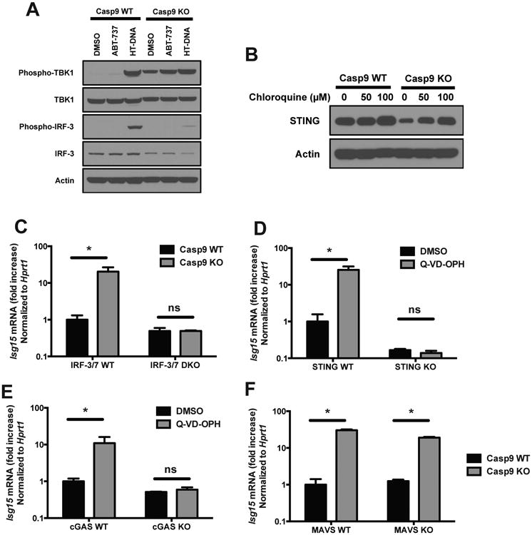
(A) Western blot analysis of the phosphorylation of TBK1 and IRF-3 in Casp9 WT and KO cells treated for 6h with vehicle (DMSO), with the Bcl-2 inhibitor ABT-737 (10 μM), or transfected with HT-DNA as a positive control (3 μg/ml, 3h). Result representative of 3 independent experiments.
(B) Western blot analysis of STING in Casp9 WT and KO cells treated for 16h with the indicated concentrations of chloroquine.
(C) Caspase-9 KO mice were crossed with IRF-3/7 DKO and the expression of ISGs in embryo heads was measured by RT-PCR. Results shown are mean ± s.d. of 3 embryos for each genotype.
(D and E) STING WT and KO primary MEFs (D) or cGAS WT and KO bone marrow-derived macrophages (E) were treated with vehicle (DMSO) or with the caspase inhibitor Q-VD-OPH (10 μM) and ISG expression was measured 48h later by RT-PCR (mean ± s.d. of triplicates, representative of 2 independent experiments).
(F) Caspase-9 KO mice were crossed with MAVS KO and the expression of ISGs in embryo heads was measured by RT-PCR. Results shown are mean ± s.d. of 2 embryos for each genotype.
*, p<0.05; ns, not significant; pairwise comparisons following two-way ANOVA.