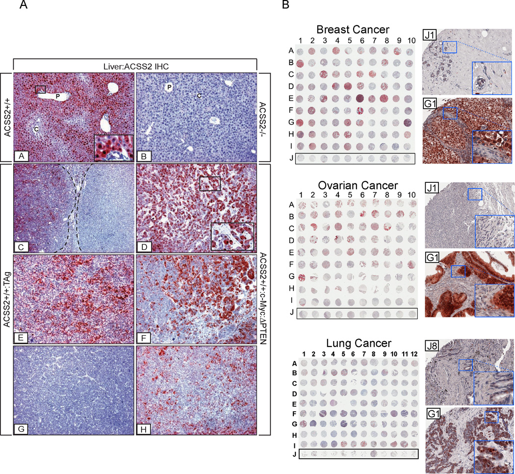
Figure 3. Immunohistochemical (IHC) Analysis of ACSS2 Expression in Tumors.
(A) Expression patterns of ACSS2 protein in normal mouse liver and liver tumors. Insets: [A] ACSS2 IHC on WT liver showing regional expression of ACSS2 across the lobule. ACSS2 is localized to the nucleus/cytoplasm in zone 1 and 2 hepatocytes, but is largely absent from zone 3 hepatocytes and biliary cells. P, portal vein; C, central vein. [B] Complete absence of ACSS2 expression in liver of an ACSS2 −/− mouse. [C], [E], [G] Variability of ACSS2 expression in tumor-laden livers of ApoE-rtTA:TRE2-TAg mice provided with 10 µg/mL doxycycline for 42–45 days. [D], [F], [H] ACSS2 expression in c-Myc:ΔPTEN tumors. AEC chromagen (red); hematoxylin counterstain (blue). [A] ×60 mag, higher mag inset ×366; [B] ×60 mag; [C] ×36 mag; [D] ×60 mag, higher mag inset ×244; [E]–[H] ×60 mag.
(B) Survey of ACSS2 expression in human breast, ovarian, and lung tumors. IHC was carried out to assess ACSS2 protein expression in a panel of human breast, ovarian, and lung tumor sections in tissue microarray format. Row J denotes samples of normal, non-cancerous tissue. For each category of tumor, a higher magnification image of one representative example of staining in normal tissue, and one representative example of staining in a high ACSS2-expressing tumor, are shown to the right. Scoring of ACSS2 expression is these TMA samples is indicated in Table S5.

