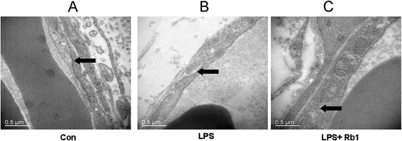
Figure 5.

Representative electron micrographs of rat pulmonary microvasculature. A: Control group; B: LPS group; C: LPS plus Ginsenoside Rb1 group. Arrow: intercellular junction. In the Con group microvasculature was lined by a layer of endothelial cells, which exhibited a rather smooth inner face with occasionally occurring vesicles in the cytoplasm. An apparent alteration occurred in the ultrastructure of the endothelial cell, characterized by the increase in the gap of intercellular junctions in LPS group.
