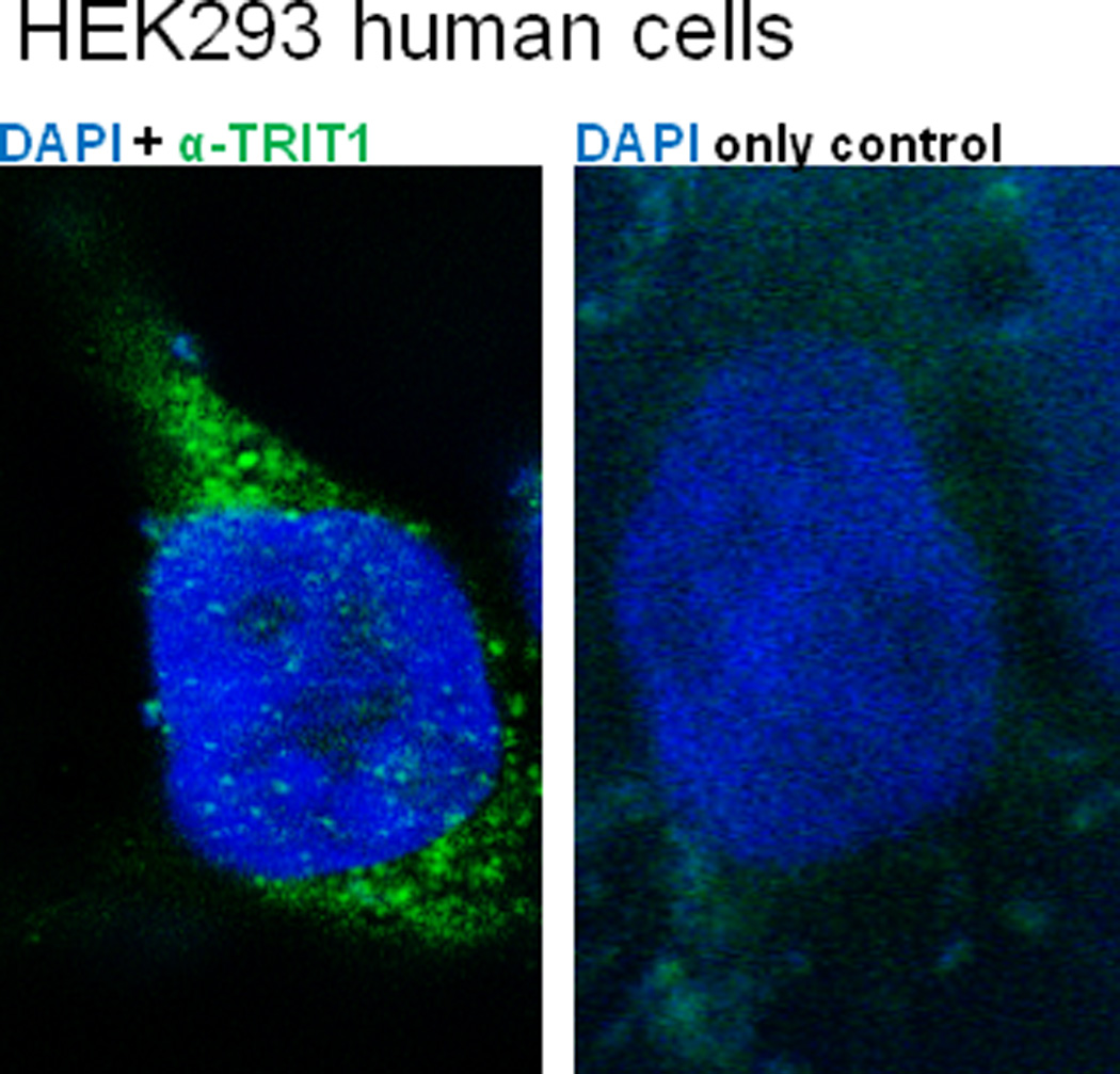
Figure 4.

TRIT1 is present in the cytoplasm and nucleus in human cells. Representative 0.25 μM slices in human cells are shown taken through the nuclei and cytoplasm from a 3D-image that was generated using AutoQuant 3D-deconvolution software. Human HEK293A cells with nuclear DAPI staining (blue). (Left panel) Cell probed with commercial TRIT1 primary antibody, with specificity verified by western blot (not shown). Signal was detected with fluorescein-conjugated secondary antibody (green). (Right panel) Cell stained with only fluorescein-conjugated secondary antibody (green) as a control. TRIT1 is primarily cytoplasmic, but there are consistently small amounts of punctate signal in optical slices through the nucleus.
