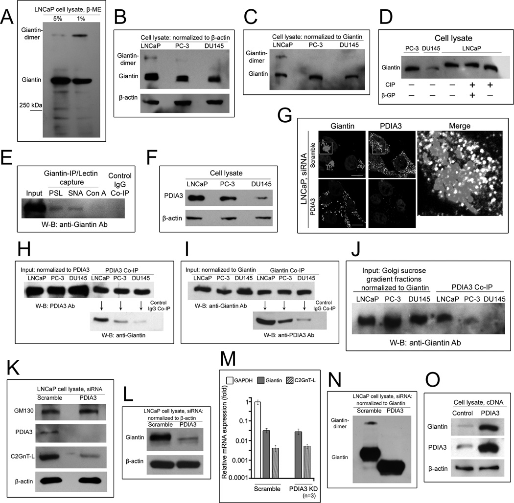
Figure 3. Giantin is a phosphorylated homo-dimer in LNCaP cells.
(A) Giantin W-B of LNCaP cell lysates prepared under high (5%) and low (1%) concentrations of β-mercaptoethanol (ME). (B, C) Giantin W-B of the cell lysates prepared under low ME. Samples were normalized to either β-actin or giantin. (D) Band-shift assay of phosphorylated giantin. The lysates of LNCaP cells were treated with CIP in the presence or absence of β-GP. (E) LNCaP cell lysate was subjected to immunoprecipitation with giantin Ab followed by pull-down using PSL, SNA, or Con A lectin. Giantin W-B of these samples shows that giantin is a glycoprotein with complex-type N-glycans. (F) PDIA3 W-B of the lysates of LNCaP, PC-3 and DU145 cells. (G) Images of giantin and PDIA3 in LNCaP cells treated with scramble or PDIA3 siRNAs. The Golgi area in the white box is enlarged and displayed as merged grey (giantin) and white (PDIA3) colors at the right side. (H, I) PDIA3 and giantin W-B of the complexes pulled down with anti-giantin and anti-PDIA3 Abs from cell lysates adjusted to equal amounts of PDIA3 (H) and giantin (I), respectively. (J) Giantin W-B of the complexes pulled down withanti-PDIA3 Ab from the Golgi fractions adjusted to equal amounts of giantin. (K, L) GM130, PDIA3, C2GnT-L and giantin W-B of the lysates of LNCaP cells treated with scramble or PDIA3 siRNAs and normalized to β-actin. (M) Quantitative real-time PCR analysis of the mRNA of giantin and C2GnT-L genes in LNCaP cells treated with scramble or PDIA3 siRNAs. (N) Giantin W-B of the lysates of LNCaP cells treated with scramble or PDIA3 siRNAs and normalized to giantin. (O) Giantin W-B of the lysates of LNCaP cells transfected with or without a full length PDIA3 cDNA. All SDS PAGE was performed with 6 or 8% gel.

