Figure 1.

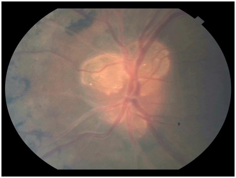
High magnification retinography of the left eye reveals several optic disc drusen, as well as bone spicule-shaped pigmentary deposits and sheathing of the retinal blood vessels.

High magnification retinography of the left eye reveals several optic disc drusen, as well as bone spicule-shaped pigmentary deposits and sheathing of the retinal blood vessels.