
Figure 2.
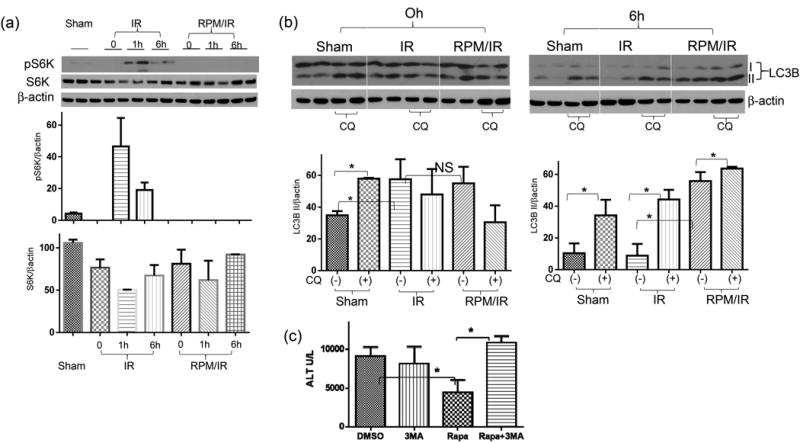
Rapamycin enhances liver autophagy during reperfusion. (a) Western blots of p70S6K in liver tissues post IR. Livers were harvested from sham or IR ones after 0, 1, 6 hrs of reperfusion (duplicate samples). Tissue protein lysates were prepared and separated by SDS-PAGE. S6K, phosphorylated S6K and β-actin levels were measured by Western blots, and protein bands were quantitated as ratios against β actin. (b) Western blots of LC3B in IR livers. Liver tissue proteins were prepared from mice after sham operation or ischemia and 0 or 6h reperfusion. To measure autophagy flux, groups of mice received CQ prior to liver ischemia, as described in the material and methods. Average LC3B II band intensities were quantitated as ratios against β actin. For tissue Western blot analysis, 2 samples/group, (c) Average serum ALT levels in mice subjected to 90m ischemia/6h reperfusion treated with vehicle (DMSO) or 3-MA, or RPM, or 3-MA/RPM prior to the start of liver ischemia, as described in the material and methods. n=4-6 mice/group.
Representative results of 2 different experiments. *p<0.05.