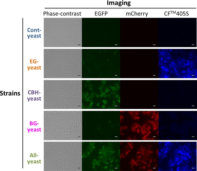
FIG 3.
Confirmation of cellulase display by immunostaining and fluorescence microscopy. Cells of 5 yeast strains were immunostained using an anti-FLAG mouse monoclonal primary antibody and CF405S-conjugated goat antimouse monoclonal secondary antibodies. Fluorescence microscopy was used to examine the fluorescence of EGFP on CBH-yeast and ALL-yeast, CF405S on EG-yeast and ALL-yeast, and mCherry on BG-yeast and ALL-yeast. Bars = 5 μm.

