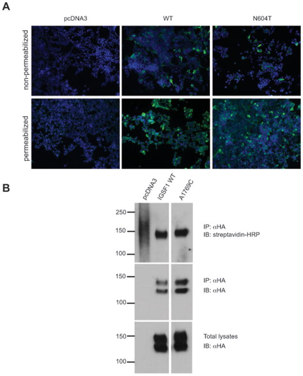
Figure 1.
IGSF1 p.N604T traffics to the plasma membrane. A – HEK293 cells were transfected with pcDNA3, IGSF1 WT or IGSF1 p.N604T constructs and subjected to immunofluorescence under permeabilizing (bottom panels) and non-permeabilizing conditions (top panels). The proteins were detected with an antibody that recognizes the N-terminus of the IGSF1 C-terminal domain. B – HEK293 cells were transfected with the indicated constructs and subjected to cell surface biotinylation followed by immunoprecipitation. Streptavidin-HRP signals of equal intensity was detected in cell lysates from WT and p.N604T transfected cells (top panel). The middle panel shows equivalent IP of the two proteins whereas the bottom panel shows equivalent expression of the two proteins. Note that IGSF1 migrates as a double on SDS-PAGE. The lower band corresponds to the core (immature) glycoform whereas the upper band is the mature glycoform.

