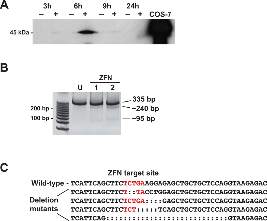
Figure 3.
ZFN-mediated mutagenesis of killifish AHR2a. A. ZFN protein expression in killifish embryos. Uninjected (−) or ZFN-injected (+) embryos were sampled at 3, 6, 9, and 24 hours after micro-injection. Homogenates were resolved on SDS-PAGE and probed with a Flag-tag antibody. Lysate from COS-7 cells transfected with the ZFN expression plasmid was run as a positive control. B. Surveyor nuclease detection of mutations in the ZFN target region of AHR2a. Each lane represents a pool of 5 embryos from which a 335 bp genomic DNA fragment was amplified. U: uninjected control, ZFN: injected embryos. Lanes 1 and 2 are two representative samples that were positive in the mutation detection assay. Approximate sizes of the Surveyor nuclease-digested fragments containing the deletions are shown (240 and 95 bp). The full-length PCR product from sample 2 was cloned and sequenced. C. AHR2a exon 3 sequence surrounding the ZFN target site (in red). Four types of deletion mutants were observed among the sequenced clones (deletions of 2, 4, 5, and 28 nucleotides).

