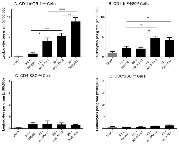
Figure 3. Blocking PD-1 ligands exacerbates innate renal inflammation after kidney IRI.
Naïve WT mice were treated with isotype control antibodies, anti-PD-L1 or anti-PD-L2 or the combination of both blocking antibodies. After 24 hours, sham or mild bilateral kidney IR surgery (24 min ischemia) was performed. At 24 hours of reperfusion kidney cell suspensions were analyzed by flow cytometry for CD45+7AAD−CD11b+GR-1high cells (A), CD45+7AAD−CD11b+F4/80int cells (B), CD45+7AAD−CD4+SSClow (C) and CD45+7AAD−CD8+SSClow (D). Absolute number of cells per gram of kidney was determined as described in the Materials and Methods section. Data are presented as mean + SEM, * denotes P<0.05; ** denotes P<0.01; *** denotes P<0.001. There are no significant differences between any group in panels C and D. N=5 for sham, 9–11 for each IRI group, pooled from 3 independent experiments.

