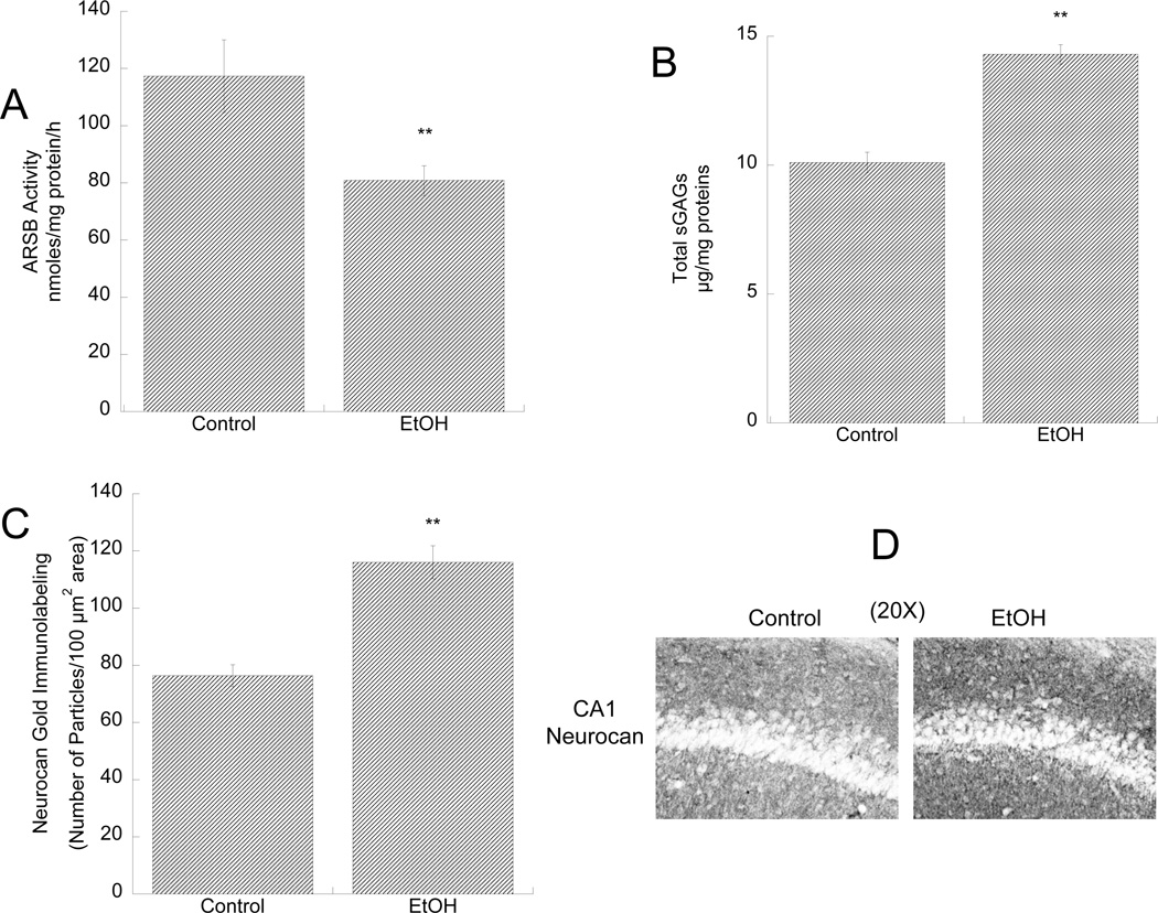
Figure 5. Effect of in vivo ethanol exposure on ARSB activity, sGAG levels, and neurocan expression in the developing hippocampus.
Male rat pups were intubated with 5g/Kg ethanol or were sham (control) intubated from PD4 to PD9. ARSB activity (A) and sGAG levels (B) were measured in hippocampus homogenates 2 h after the last intubation. **: p< 0.01; by the Student’s t-test (n=3). C, D: Neurocan expression in the stratum oriens of the CA1 region of the hippocampus was determined by gold-immunolabeling histochemistry followed by quantification using the Loats Image Analysis System. C: Quantification of neurocan gold-immunoparticles expressed as number of immunogold particles/100 µm2 area. **: p<0.01; by the Student’s t-test; n=3. D: Representative images of neurocan gold-immunolabeling staining in control (left image) and ethanol-treated (right image) pups.

