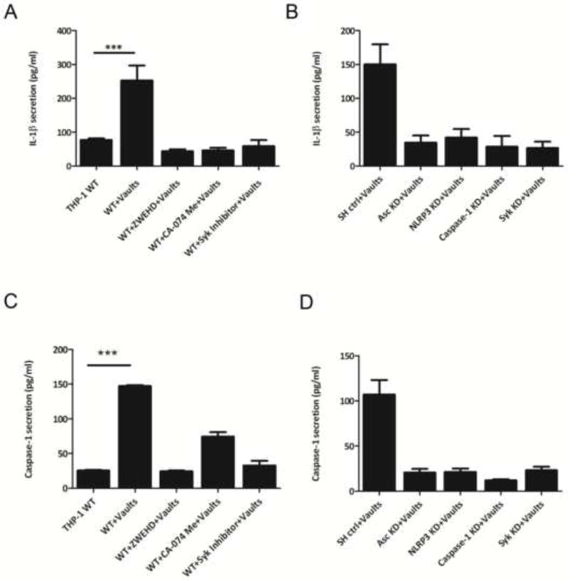
Figure 1. PmpG-1-vaults activate the NLRP3 inflammasome and induce IL-1β secretion, as measured by an ELISA assay.

THP-1 (1×10⁶) wild type (WT) cells (A) were incubated in 6-well plates with RPMI 1640 media. Inhibitors of caspase-1 (ZWEHD) or cathepsin B (CA-074) were added individually 1.5 h prior to incubation with PmpG-1-vaults, and the Syk inhibitor was added 0.5 h prior to incubation with PmpG-1-vaults. THP-1 knockdown (KD) cells (B) were incubated with media alone, and 500 μg of PmpG-1-vaults were added to each well, except the WT control. Culture supernatants were collected 6 hrs post-incubation and IL-1β was measured by ELISA. IL-1β levels secreted by cells with inhibitors vs cells without inhibitors and by WT vs KD cells was significantly different (p<0.001). (C) THP-1 (1×10⁶) WT cells were incubated in 6-well plates. ZWEHD or CA-074 was added individually 1.5 h prior to incubation with PmpG-1-vaults, and the Syk inhibitor was added 0.5 h prior to incubation with PmpG-1-vaults. (D) THP-1 knockdown (KD) cells were incubated with media alone, and 500 μg of PmpG-1-vaults were added to each well, except the WT control. Culture supernatants were collected 6 hrs post-incubation and caspase-1 was measured by ELISA kit. The mean ±SD of a representative experiment from six times was analyzed by ANOVA. Caspase-1 levels secreted by cells with inhibitors vs cells without inhibitors and by WT vs KD cells was significantly different (p<0.001). In all cases, cell supernatants were measured in triplicate.
