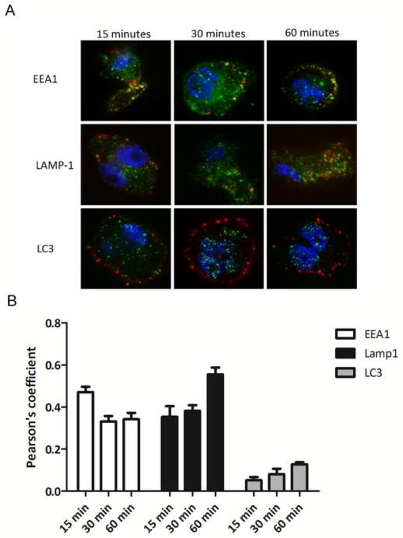
Figure 5. Uptake of PmpG-1-vaults and colocalization within the endocytic pathway.

(A) THP-1 cells were grown on 18 mm glass cover slips and treated with 30 ug of DyLight 650 labeled PmpG-1-vaults for 15, 30, and 60 min and imaged by confocal microscopy. For immunofluorescence staining, THP-1 cells were reacted with either anti-EEA1 mouse mAb, anti-Lamp1 mouse mAb, or anti-LC3 mouse mAb followed by Alexa Fluor 488-conjugated goat anti-mouse to identify endocytic compartments. (B) Colocalization of PmpG-1-vaults within each compartment was determined by calculation of the Pearson’s correlation coefficient of the red and green channels using ImageJ.
