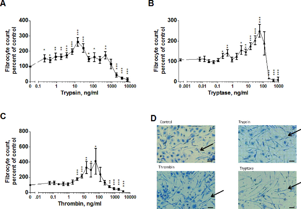
Figure 1. Trypsin, tryptase and thrombin potentiate fibrocyte differentiation.
PBMC were cultured in serum free media in the presence of the indicated concentrations of (A) trypsin (n=13), (B) tryptase (n=15), or (C) thrombin (n=14) for 5 days. Fibrocyte counts were normalized for each donor to the no-protease control. The no-protease controls developed 767 ± 90 fibrocytes per 105 PBMC. Values in A through C are mean ± SEM; the absence of error bars indicates that the error was smaller than the plot symbol. * indicates p < .05, ** p < .01, and *** p < .001 compared to the no-protease control (t-test). (D) Images of PBMC after 5 days with no protease (control), 12.5 ng/ml trypsin, 12.5 ng/ml thrombin, or 55 ng/ml tryptase. Arrows indicate fibrocytes. Bar is 40 µm.

