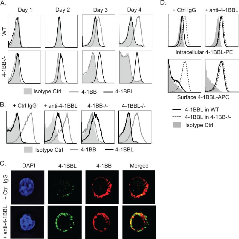
Figure 1. Expression of 4-1BB and 4-1BBL on activated T cells.
WT, 4-1BB−/−, and 4-1BBL−/− naive OT-II CD4 T cells were either activated with WT DCs and 1μM OVA peptide for varying lengths of time (A) or anti-CD3 and anti-CD28 in the presence of soluble anti-4-1BBL (19H3) or Ctrl IgG for 48 hrs (B-D). (A and B) 4-1BB and 4-1BBL surface expression in gated CD44hi Vα2+Vβ5+ T cells by flow cytometry. (C) 4-1BB and 4-1BBL expression in T cells from (B) analyzed by confocal microscopy. Green, 4-1BBL; red, 4-1BB. (D) Intracellular staining for 4-1BBL (top) after surface 4-1BBL was stained with saturating amounts of the same detection Ab conjugated to a different dye. Data are representative of three to five independent experiments.

