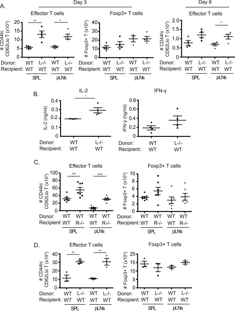
Figure 5. 4-1BBL limits T cell activation in vivo under non-inflammatory conditions.
(A) Sorted naïve WT or 4-1BBL−/− (L−/−) Ly5.2+ OT-II T cells (2 x 10⁶) were adoptively transferred into WT Ly5.1+ congenic recipient mice. One day later, mice were immunized i.v. with 5μg of OVA peptide (323-339) in PBS. After 3 days (left and middle) or 6 days (right), the number of effector (CD44hi CD62Llo) or Foxp3+ OT-II (Vα2+Vβ5+Ly5.2+) T cells was calculated in spleens (SPL) and peripheral lymph nodes (pLNs). (B) Splenocytes from (A), taken at day 3, were stimulated with PMA (5ng/ml) and ionomycin (500ng/ml) for 5 hr, and IL-2 and IFN-γ production were measured by ELISA. (C) Sorted naïve Ly5.1+ congenic WT OT-II T cells were transferred into WT or 4-1BB−/− (R−/−) Ly5.2+ recipients. Mice were immunized and analyzed at day 3 as in (A). (D) Recipients of 4-1BBL−/− T cells were challenged with 25μg OVA peptide in PBS similar to (A) and then re-challenged with 10μg OVA peptide 4 days later. Accumulation of effector and Foxp3+ OT-II T cells was analyzed after a further 3 days. All data show numbers of T cells or amounts of each cytokine in individual recipient mice, with means ± sem for each group, and are representative of at least two independent experiments in each case.

