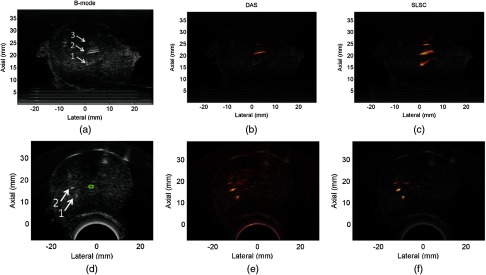
Fig. 4.

(a) Linear and (d) curvilinear ultrasound images of the ex vivo prostate implanted with the three seeds shown in Fig. 2. The arrows point to the visible seeds in the B-mode images, while the urethra is contoured in green. (b,e) Corresponding linear and curvilinear DAS photoacoustic images of the seeds overlaid on the ultrasound images. (c,f) Corresponding linear and curvilinear SLSC photoacoustic images overlaid on the ultrasound images. A transurethral light delivery method was utilized to obtain the photoacoustic images.
