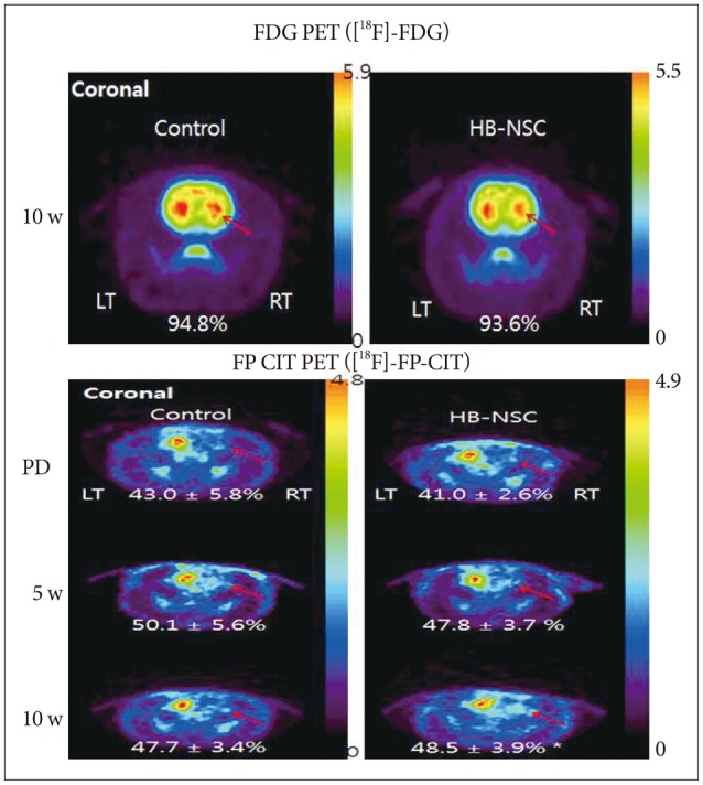
Fig. 2.

Coronal images of [18F]-FDG and [18F]-FP-CIT PET scans in striatal area are shown, respectively. In the [18F]-FDG PET scan 10 week posttransplantation, the uptake did not show significant differences in bilateral striatum in control group as well as HB-NSC group, which means PD models were created successfully. [18F]-FP-CIT PET scans were performed before transplantation (PD), 5 weeks after transplantation (5 w), and 10 weeks after transplantation (10 w). The HB-NSC group showed a statistically significant recovery in the uptake of [18F]-FP-CIT in the transplanted striatum (arrow) at 10 weeks posttransplantation compared to PD (*p< 0.05) whereas there was no significant improvement of uptake at 5 weeks posttransplantation (p=0.065), suggesting that dopamine is secreted by the transplanted cells. The color bar indicates the relative uptake intensity and shows maximum standardized uptake values in the image.
