
Fig. 3.
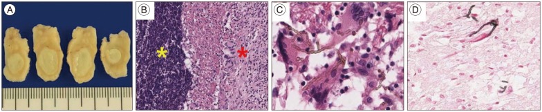
Gross preparation shows white, well-demarcated, round masses in brain parenchyma (A). Hematoxylin and eosin stain reveals necrotizing granulomas (red stared, ×100) and inflammatory infiltrates (yellow stared, ×100) (B), as well as brown colored septated hyphae (×1000) (C). Black colored melanin pigments are present in branched fungal hyphae on Fontana-Masson stain (×400) (D).