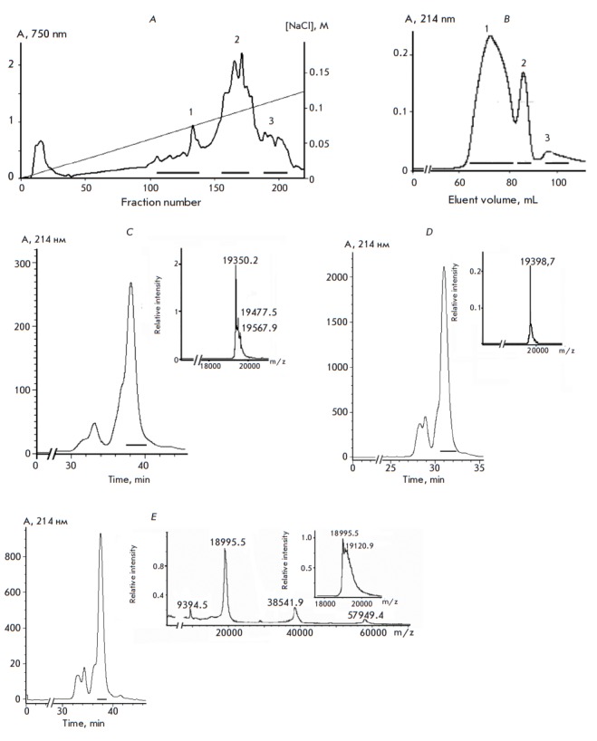
Fig. 1.

Chromatography of the hemolytic active polypeptides of the sea anemone, H. crispa. A – Ion exchange chromatography of the total protein preparation on a CM- 32 cellulose column. Boundaries for combining fractions of polypeptides with hemolytic activity are denoted. B – FPLC gel filtration of fraction 2 polypeptides after ion exchange chromatography on a Superdex Peptide 10/30 column; boundaries for combining fractions are denoted. C–E – RP-HPLC of fractions 1–3 polypeptides, after gel filtration, on a Nucleosil C18 column and mass spectra of the obtained compounds
