
Figure 3.
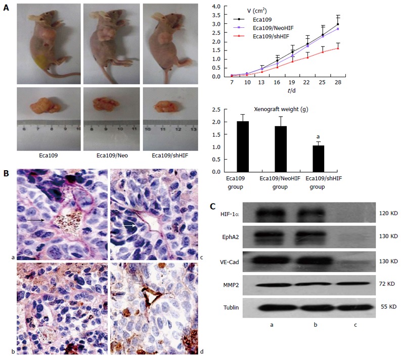
In vivo effects of hypoxia inducible factor-1α knockdown on regulation of esophageal cancer cell xenograft formation and gene expression. A: Tumor size curve and weight of xenograft (Eca109/shHIF-1α cells vs Eca109 cells and Eca109/HIF-1α Neo cells, aP < 0.05); B: VM structure (arrow) in xenografts of three groups (a: Eca109 group; b: Eca109/shHIF group; c: Eca109/NeoHIF group; d: Normal vessels; arrow, VM structure); C: Western blot detected HIF-1α and EphA2, VE-cadherin and MMP2 expression (a: Eca109 group; b: Eca109/NeoHIF group; c: Eca109/shHIF group; Eca109/shHIF-1α cells vs Eca109 and Eca109 HIF-1α Neo control cells, P < 0.05). HIF: Hypoxia inducible factor; VM: Vasculogenic mimicry.