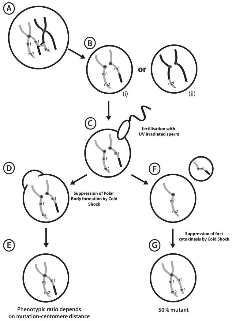
Figure 1. Formation of Haploid and Gynogenetic Embryos.
(A) Diplotene oocyte in a female hybrid for mutagenized gray strain chromosomes and polymorphic black strain, showing a crossover event between mutant loci m1 and m2. (B) Unfertilized eggs showing segregation of sister chromatids after Meiosis I. Note that regions where centromeres hold sister chromatids together are homozygous. (C) UV-irradiated sperm activates development without paternal genetic contribution, forming haploid embryos (F) following polar body extrusion. (D) Early cold shock suppresses formation of the second polar body, with the resulting gynogenote (E) rescued to diploidy and retaining both sets of sister chromatids. Recessive phenotypes at loci closer to centromeres (m1) are more likely to be uncovered than distal loci (m2), where recombination produces heterozygous wild type. (G) Late Cold Shock of haploid embryos following DNA replication prevents first cytokinesis, rescuing haploid to completely homozygous diploids.

