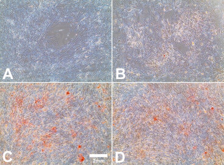
Figure 6.
Influence of Ag-NP/ Ag+ ions on hMSCs during osteogenic differentiation. After 21 d of cell culture (bright-field images), staining with alizarin red S was used to visualize calcium accretion in cells cultured under osteogenic conditions. hMSCs incubated in the presence of osteogenic-differentiation media served as the positive control (C). hMSCs incubated in the presence of RPMI/FCS served as the negative control (A). hMSCs incubated with 10 µg·mL−1 Ag-NP (B) or with 1.0 µg·mL−1 Ag+ ions (D) for 24 h, and followed by osteogenic-differentiation media for a further 21 d.

