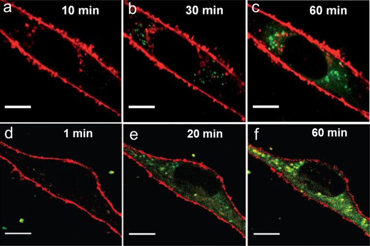
Figure 4.
Two-color merged confocal images of live human MSCs exposed to NPs (green) in PBS for different times. (a–c) PS− NPs, 75 µg/mL; (d–f) PS+ NPs, 7.5 µg/mL. Scale bar, 10 µm. Cell membranes (in red) are stained with CellMask DeepRed. Images in panels a–c were reproduced with permission from [32]. Copyright 2011 Royal Society of Chemistry. Images in panels d–f were reproduced with permission from [33]. Copyright 2010 American Chemical Society.

