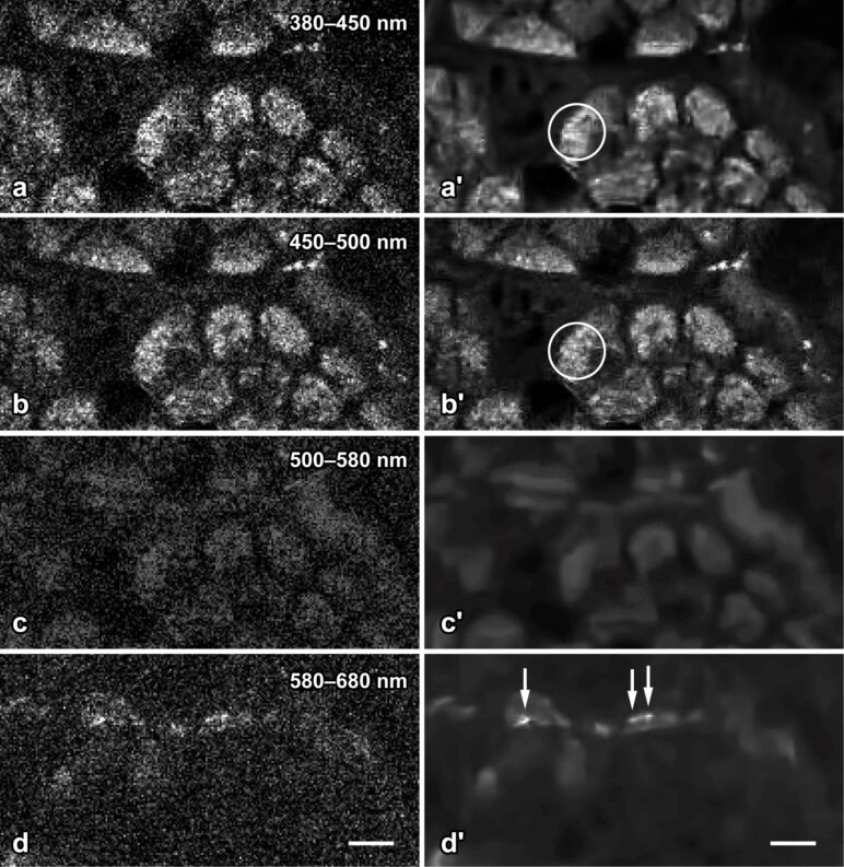
Figure 7.
Epithelial cells and quantum dot nanoparticles of the murine gut mucosa in intravital 2-photon microscopy. The eight images correspond to the same field of view; raw data are shown in the left column (a–d), and the corresponding denoised images in the right (a′–d′). Nonlinear excitation of tissue and nanoparticle fluorescence was carried out 730 nm. The emitted light was split to four spectral channels, separated by dicroic mirrors at 450, 500 and 580 nm. The modified BM3D algorithm successfully reduces shot noise, but preserves fine structural details in the apical cytoplasm of the cells (encircled in a′ and b′). Quantum dot nanoparticles (arrows in d′) adhere to the apical surface of the cells and emit in channel 4 only. Denoising by the modified BM3D algorithm considerably facilitates the perception of the nanoparticles by the human observer (compare d to d′) and allows for automated image analysis to be applied to denoised 2PM images. Bar = 5 µm.

