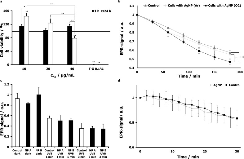
Figure 4.
Biological responses of skin tissue and skin cells to particle exposure. The viability of HaCaT cells after 1 h and 24 h incubation with AgNP at different concentrations was assessed by using the XTT assay (a). HaCaT cells were incubated with 30 µg/mL of AgNP produced and stored under ambient air conditions or in argon atmosphere, respectively, and investigated by means of EPR spectroscopy. The used spin marker TEMPO (5 µM) becomes EPR-invisible when reacting with ROS (b). In order to analyze ROS production in whole skin, the EPR-signal intensity was monitored after the application of TiO2 on porcine ear samples at two different concentrations: 40 mg/mL (NPs A), 400 mg/mL (NPs B) and after irradiation after 1 or 3 min UVB light (210 and 630 mJ/cm2, respectively) and respective controls (c). Similarly, the EPR signal of porcine skin was followed after the topical application of AgNP (0.446 mg/mL) for 1 h. Control samples were treated with PBS only (d).

