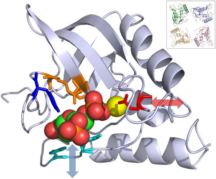
Fig. 2.
Active-site conformational dynamics in crystalline staphylococcal nuclease. The backbone is rendered using a ribbon, and the P41 unit cell packing along the c axis is shown in the Inset (the screw axis translation is into the page, with the green copy closest and the orange copy farthest away). The residues are shown using sticks, proceeding counterclockwise: Glu43 (red), Arg35 (orange on the β-sheet), Arg87 (orange on the loop), Tyr85 (blue), Tyr115 (cyan), and Tyr113 (cyan). The rest of the protein is rendered as a gray cartoon. The inhibitor thymidine 3′,5′-bisphosphate is shown using spheres and the calcium ion using a yellow sphere. Arrows indicate the direction of motion in the two dominant principal components of the microsecond MD simulation. The loop containing Glu43 (red) moves in the approximate direction indicated by the transparent red double-headed arrow. The loop containing Tyr113 and Tyr115 (cyan) moves in the approximate direction indicated by the transparent blue double-headed arrow. The region containing Tyr85 (blue) and Arg35 and Arg87 (orange) moves much less by comparison. The image was created using PyMOL (66).

