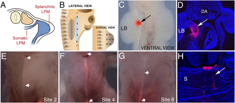
Fig. 2.
The sternum precursor cells reside in the LPM, ventral to the forelimb bud. (A) Schematic of transverse section through HH20 chick showing LPM subdivision into somatic and splanchnic domains. (B) Schematic of DiI injection sites and adjacent somites with sternum precursor population highlighted (blue). Ventral whole-mount view (C) and transverse section of HH20 embryos showing DiI-labeling (arrows) (D) following injection into site 4. Limb bud is labeled LB; dorsal aorta is labeled DA. (E–G) Ventral whole-mount view of harvested, skinned HH36 embryos showing DiI-labeled cells at the midline (boundaries of population shown by white arrowheads) following injection into HH20 embryos at sites 2, 4, and 6, respectively. (H) Transverse section through a harvested embryo showing DiI-labeling in the sternum (arrow). S, sternum.

