Fig. 2.
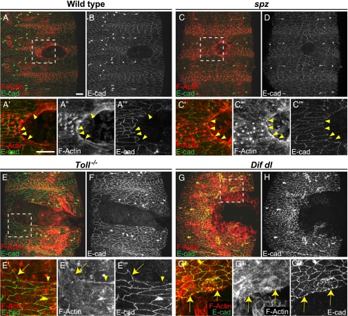
Wounded Toll−/− mutants and Dif dl double mutants do not build a continuous actin cable. Embryos of stage 15 were stained for F-actin (phalloidin) and E-cadherin at 2 h after wounding. (A, C, E, and G) are merged images, whereas (B, D, F, and H) show only E-cadherin. Identical settings were used for all images. Areas for the close-ups are outlined by squares in A, C, E, and G. (A, A′, A′′, A′′′, and B) In wild type, wound-edge cells are contracting and the wound is closing. Wound-edge epidermal cells show a continuous actin cable (A′ and A′′). E-cadherin (B and A′′′, arrowheads) is excluded from the membranes facing the wound. (C, C′, C′′, C′′′, and D) In spz embryos, wound-edge cells are contracting and wound closure is similar to wild type. Wound-edge cells have a continuous actin cable (C′ and C′′). E-cadherin (D and C′′′, arrowheads) is not present at the membranes that face the wound. (E, E′, E′′, E′′′, and F) In Toll−/− embryos, the actin cable is discontinuous. Epidermal cells that maintain high levels of E-cadherin at the membranes bordering the gap (E′′′, arrow) do not assemble an actin cable (E′ and E′′, arrow). In wound-edge cells where some actin bundles are assembled (E′ and E′′, arrowhead), E-cadherin is reduced at the membranes facing the gap (E′′′, arrowhead). (G, G′, G′′, G′′′, and H) In Dif dl embryos, the actin cable is discontinuous. Epidermal cells that maintain high levels of E-cadherin at the membranes bordering the gap (G′′′, arrows) do not assemble an actin cable (G′ and G′′, arrows) at the wound margin. (Scale bars: 10 μm.) Images are the maximum z projections of 11.1- to 22-μm stacks (30–55 slices). Close-ups are the maximum z projections of 5.2- to 18.4-μm stacks (14–46 slices).

