Fig. 1.
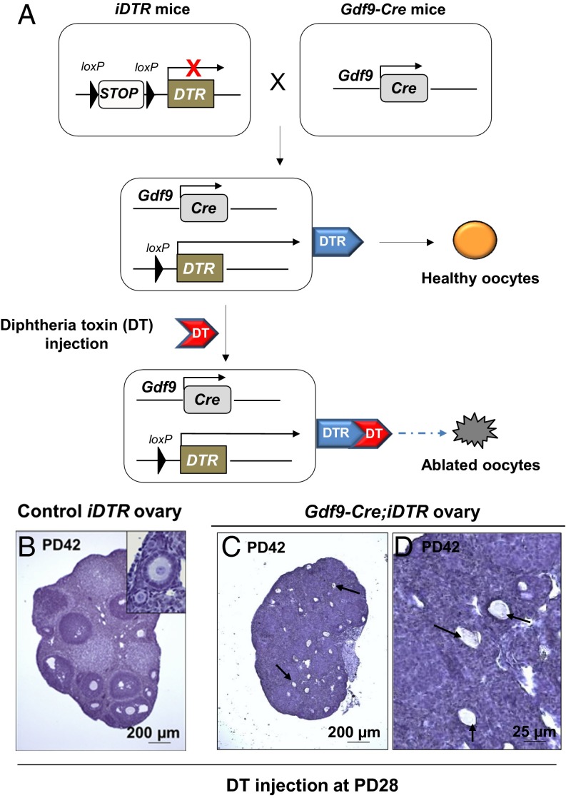
Specific and efficient ablation of oocytes in the Gdf9-Cre;iDTR mouse ovaries. (A) Illustration of the diphtheria toxin (DT)-induced ablation of oocytes in Gdf9-Cre;iDTR mouse ovaries. In Gdf9-expressing oocytes, the Cre recombinase removes the loxP-flanked STOP sequence and allows the expression of the DTR. Upon DT administration, the Gdf9-expressing oocytes are selectively ablated. (B–D) All existing oocytes were ablated in Gdf9-Cre;iDTR mouse ovaries after DT injection. DT was given to PD28 females for 5 consecutive days, and ovaries were examined 2 wk later. Compared with the normal ovarian development in iDTR females (B), Gdf9-Cre;iDTR ovaries demonstrated a complete loss of all oocytes (C and D). Only follicle structures without oocytes or with oocyte debris were observed (C and D, arrows) (n = 6).

