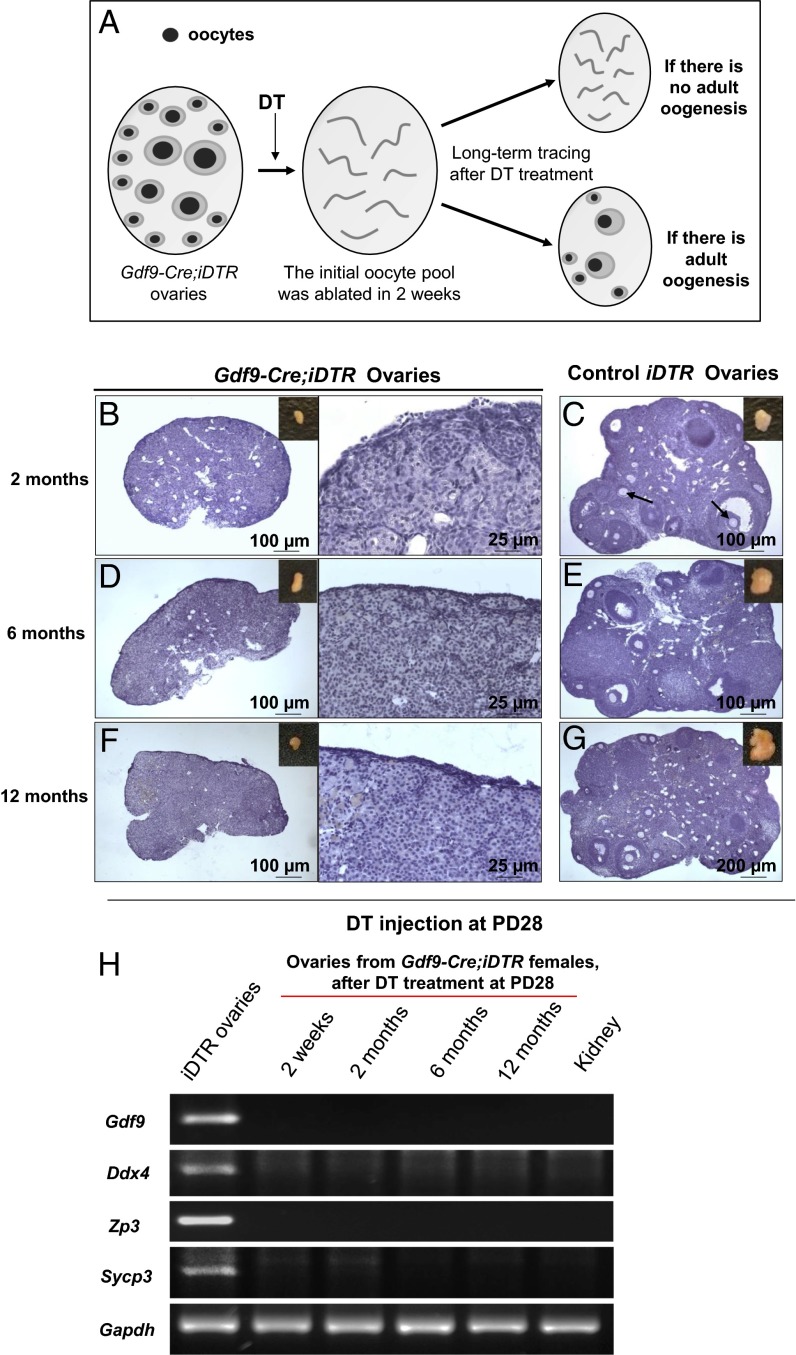
Fig. 2.
No oocyte regeneration occurred in oocyte-ablated Gdf9-Cre;iDTR mouse ovaries. (A) Schema for long-term tracing of oocyte regeneration in Gdf9-Cre;iDTR mouse ovaries. Neogenic oocytes and follicles should be visible in the ovaries of DT-injected mice if there is oocyte regeneration during the 12-mo tracing study. (B, D, and F) No live oocytes or follicles were observed in Gdf9-Cre;iDTR ovaries at 2 mo (n = 6) (B), 6 mo (n = 6) (D), or 12 mo (n = 6) (F) after DT administration. (C, E, and G) As controls, the ovarian morphology of iDTR females was normal at 2 mo (n = 4) (C), 6 mo (n = 6) (E), and 12 mo (n = 6) (G) after DT administration. (H) Comparative gene expression profiling of germ-cell markers in oocyte-ablated Gdf9-Cre;iDTR ovaries. Semiquantitative RT-PCR analysis showed that none of the germ-cell markers studied, including Gdf9, Ddx4, Zp3 and Sycp3, were expressed in the Gdf9-Cre;iDTR ovaries from 2 wk to 12 mo after DT administration. An RNA sample from the mouse kidney was used as a negative control, and Gapdh was used as the internal loading control. All experiments were repeated at least three times, and representative images are shown.

