Fig. 3.
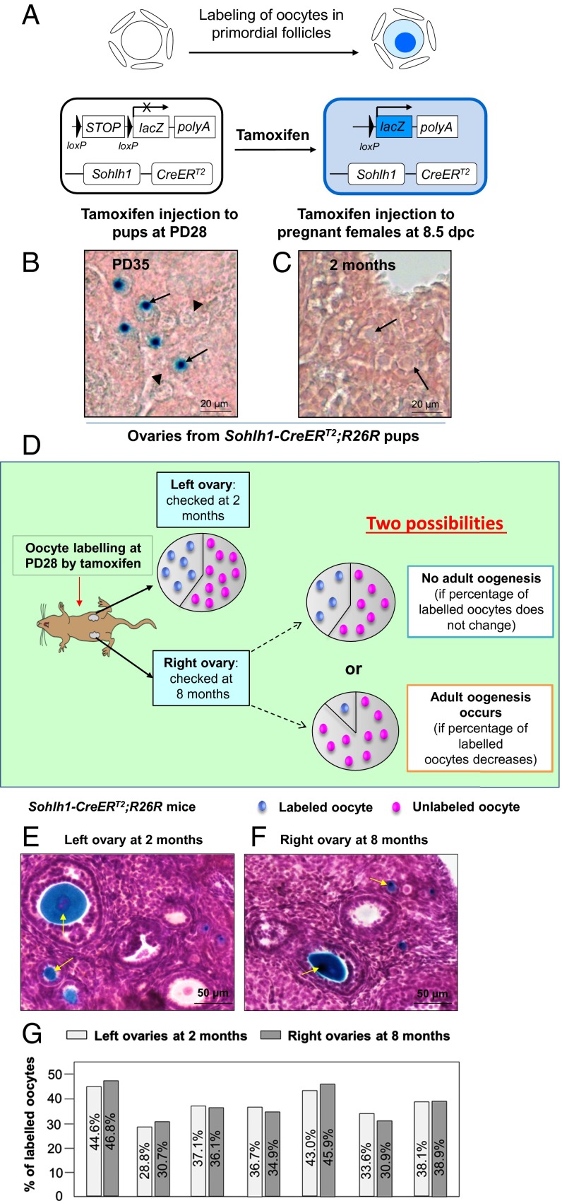
The developing oocytes in later reproductive life originated from immature oocytes that were labeled in early life in Sohlh1-CreERT2;R26R females. (A) Illustration of the tamoxifen-induced labeling of oocytes in primordial follicles of the Sohlh1-CreERT2;R26R ovary. CreERT2 recombinase is expressed in Sohlh1-expressing oocytes of primordial follicles. Upon tamoxifen administration, the CreERT2 recombinase removes the STOP sequence and turns on the expression of lacZ (the gene encoding β-galactosidase), and this generates a blue color in the oocyte cytoplasm after β-galactosidase staining. (B) Labeling oocytes of primordial follicles by postnatal tamoxifen injection. Tamoxifen was given to Sohlh1-CreERT2;R26R females for 7 consecutive days at PD28, and ovaries were collected for analysis at PD35 (n = 6). The labeled oocytes were visualized by their blue color after β-galactosidase staining (arrows) whereas the unlabeled oocytes showed only background color (arrowheads). (C) No PGCs were labeled by embryonic tamoxifen injection. R26R females were plugged with Sohlh1-CreERT2 males, and tamoxifen was given to pregnant females from 8.5 to 12.5 dpc. No labeled oocytes were observed at 2 mo in the ovaries of Sohlh1-CreERT2;R26R pups that were born from the tamoxifen-injected females (n = 6). (D) Schema for long-term tracing of oocyte regeneration in Sohlh1-CreERT2;R26R mouse ovaries. The ratio of labeled-to-unlabeled oocytes should remain the same if there is no adult oogenesis but should decrease if oocyte neogenesis occurs. (E and F) Labeling the existing oocytes in the adult mouse ovary. The labeled oocytes (identified by the blue dots) can be observed in both the left ovary at 2 mo (E) and the right ovary at 8 mo (F) after tamoxifen injection. Some labeled oocytes had entered into the growing phase (arrows). (G) The ratio of labeled-to-unlabeled oocytes remained the same in each mouse at 2 mo (left ovaries) (n = 7) and at 8 mo (right ovaries) (n = 7) after tamoxifen injection. The similarity in the proportion of labeled oocytes indicates that no oocyte neogenesis occurred in the adult mouse ovaries.

