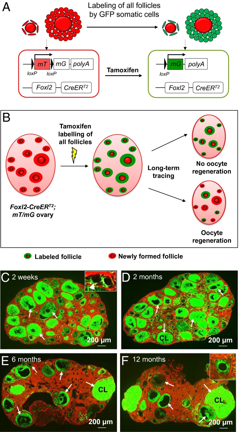
Fig. 4.
Long-term tracing of follicle populations by labeling the follicular somatic cells in adult Foxl2-CreERT2;mT/mG ovaries. (A) Illustration of the tamoxifen-induced labeling of follicles in Foxl2-CreERT2;mT/mG mice. In Foxl2-expressing follicular somatic cells, the CreERT2 recombinase is not active and the cells express mT, a red fluorescent protein. Upon tamoxifen injection, the CreERT2 recombinase deletes the mT region and switches on the expression of mG, a green fluorescent protein. Thus, the Foxl2-expressing follicular somatic cells were labeled with green fluorescence. (B) Schema for long-term tracing of oocyte regeneration in Foxl2-CreERT2;mT/mG mouse ovaries. All follicular somatic cells were labeled with the green fluorescent mG at PD28. Follicles with only red (mT) somatic cells would be observed if oocyte regeneration occurred during the tracing study. (C–F) The mice were given an i.p. injection of 80 mg⋅kg−1 BW (∼2 mg per mouse) tamoxifen per day for 7 consecutive days starting at PD28. The mice were killed at 2 wk (n = 6) (C), 2 mo (n = 6) (D), 6 mo (n = 6) (E), and 12 mo (n = 6) (F) after the injection. Ovaries were collected and all sections were carefully checked under the microscope, and a follicle or a corpus luteum was considered to be labeled if green fluorescent granulosa or luteal cells were observed. At 2 wk after tamoxifen injection, all follicles, including the dormant primordial follicles in the cortex (C, Inset, arrowhead) and growing follicles in the medulla (C, arrows), were labeled with green fluorescence in their somatic cells. All follicles (D–F, arrows) or corpora lutea (D–F, CL) were labeled with green fluorescence in their somatic cells, and no follicles with only red somatic cells were found in mice killed at 2 mo (D), 6 mo (E), or 12 mo (F) after tamoxifen injection.

