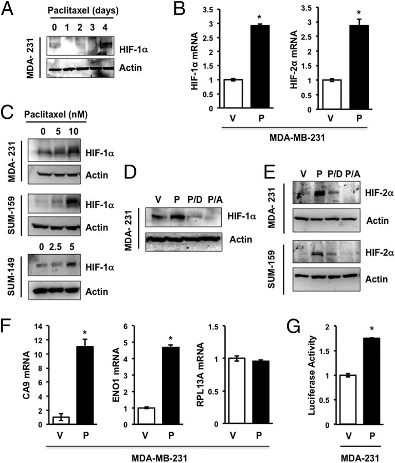
Fig. 1.
Paclitaxel induces HIF expression and signaling. (A) Immunoblot assays were performed to analyze HIF-1α and actin expression following exposure of MDA-MB-231 (MDA-231) cells to 10 nM paclitaxel for 1–4 d or vehicle (0 d). (B) MDA-231 cells were treated with vehicle (V) or 10 nM paclitaxel (P) for 4 d and aliquots of total RNA were assayed by RT-qPCR using primers specific for HIF-1α or HIF-2α mRNA relative to 18S rRNA, and the results were normalized to cells treated with V (mean ± SEM; n = 3). *P < 0.001 by Student's t test. (C) MDA-231 and SUM-159 cells were treated with 0–10 nM paclitaxel, whereas SUM-149 cells were treated with 0–5 nM paclitaxel for 4 d, and immunoblot assays of HIF-1α and actin were performed. (D and E) Cells were treated with vehicle (V) or 10 nM paclitaxel, either alone (P) or in combination with 100 nM digoxin (P/D) or 1 µM acriflavine (P/A), for 4 d, and the expression of actin, HIF-1α (D) and HIF-2α (E) was determined by immunoblot assays. (F) RT-qPCR was performed as described above to assay CA9, ENO1, or RPL13A mRNA (mean ± SEM; n = 3). *P < 0.001 by Student's t test. (G) MDA-MB-231 cells were transfected with HIF-dependent firefly luciferase reporter p2.1 and control Renilla luciferase reporter pSV-RL, then exposed to vehicle control (V) or 10 nM paclitaxel (P) for 4 d, and the ratio of firefly:Renilla luciferase activity was determined. *P < 0.0001 by Student's t test.

