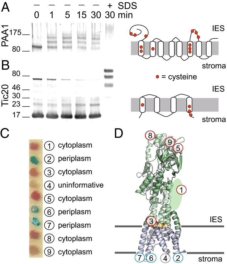
Fig. 5.
Topology and orientation of PAA1. PEG-Mal labeling of P. sativum inner envelope vesicles in the presence or absence of 1% SDS was performed for the indicated time subsequent to detection of pea PAA1 (A) and Tic20 (B) by immunoblotting. The location of cysteines in each protein is shown to the right. (C) E. coli cells expressing various translational fusions between the PhoA-LacZα reporter cassette and AtPAA1 were plated on dual indicator plates, which contain chromogenic substrates. Hydrolysis of 5-bromo-4-chloro-3-indolyl phosphate by PhoA yields a blue color, whereas hydrolysis of 6-chloro-3-indolyl-β-d-galactoside by LacZ yields a red color, allowing assignment of the PhoA-LacZα reporter to either the periplasmic space or cytoplasm, respectively. (D) Location of each fusion construct from C is overlaid on the structural model of AtPAA1.

