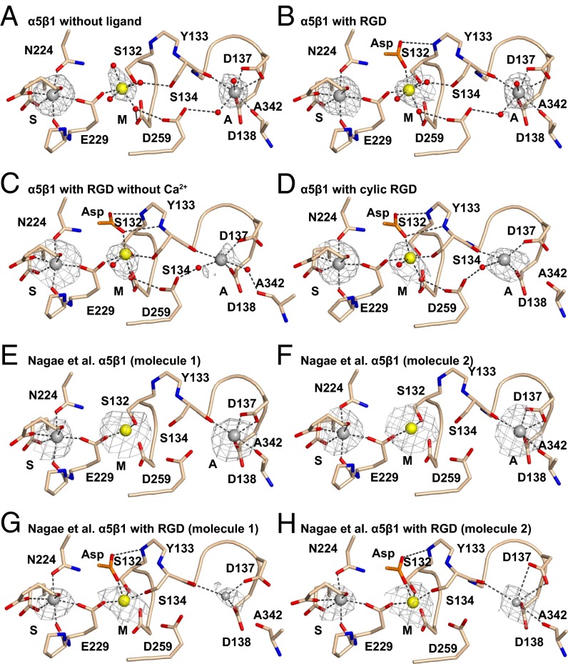
Fig. 2.
βI domain metal binding sites. (A–H) The indicated structures are in identical orientations. Backbones and side chains of metal-coordinating residues are colored wheat with red oxygens and blue nitrogens. Waters are shown as small red spheres. Ca2+ (silver) and Mg2+ (gold) are shown as large spheres. Putative ADMIDAS Ca2+ ions not included in molecular models in G and H are shown as small spheres. Simulated-annealing omit map Fo − Fc electron density contoured at 2.5σ is shown as mesh around metal ions.

