Fig. 2.
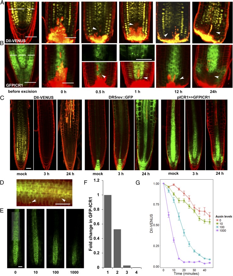
High auxin concentrations initiate rapid GFP-ICR1 decay. Roots expressing (A) DII-VENUS or (B) GFP-ICR1 were sectioned 170 μm from the tip (dashed line). After 30 min, GFP-ICR1 started to appear in intracellular compartments and then disappeared concomitant with reduction in the DII-VENUS signal. Arrowheads denote (A) reduced DII-VENUS fluorescence and (B) intracellular GFP-ICR1 compartments and reduced fluorescence. (C) Incubation in 0.5 μM NAA led to rapid decay of the DII-VENUS signal, induction of DR5rev::GFP, and decay of GFP-ICR1 500 μm from the root tip. (D) GFP-ICR1 was detected in intracellular compartments ∼500 μm above the root tip after 4 h of incubation with 10 μM IAA (arrowheads). (E) Dose-dependent destabilization of GFP-ICR1 in seedlings incubated for 20 h in increasing concentrations of IAA (0–1,000 nM). (F) Quantification of the GFP-ICR1 signal shown in E. (G) DII-VENUS signal decreased after incubation of seedlings in increasing IAA concentrations (in nanomolar). Error bars are SE. (Scale bars: 50 μm.)

