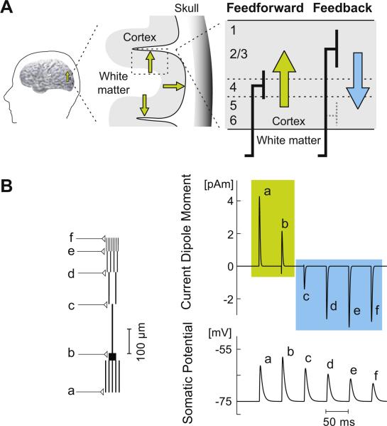
Figure 2. Dependence of the current dipole direction on the spatial location of synaptic input.
A. Schematic illustration of the laminar distribution of feedforward and feedback synaptic input (thick lines) into a cortical area (adapted from [37]). The hypothesized direction of the resulting macroscopic current dipole is outward, i.e., away from the white matter, for feedforward input (green arrow) and inward for feedback input (blue arrow). The cross-section of a region of the cortex on the left illustrates three dipoles (small green arrows) with different physical orientations: the two dipoles depicted in the sulcal walls are tangential with respect to the skull, the middle one in the gyrus is radial; however, the physiological direction for all of these is outwards. Note that in Fig. 1B the dipoles and the cortical lamina are “upside down” compared with this laminar diagram, because those sources are located in the inferior surface of the occipitotemporal cortex.
B. A simple computational model of a pyramidal neuron to demonstrate how the direction of the current dipole can depend on the location of synaptic input. The compartmental model, built using the NEURON software (www.neuron.yale.edu/neuron/), consisted of cylindrical segments representing the soma, five basal dendrites, and an apical dendrite with a trunk and three levels of bifurcations (left). All segments were assumed to be oriented perpendicular to the cortical surface. Passive membrane properties were assumed [38]. Synaptic inputs were modeled as changes in the transmembrane conductivity in the form of the alpha function with time constant τ = 0.7 ms. The synapses were placed at the end points of the dendritic segments (labeled “a” to “f”) and activated one at a time. The net current dipole was calculated by summing the dipole moment, obtained by multiplying the intracellular axial current by the length of the segment, for each segment of the pyramidal cell model [30]. When excitatory input was located at a basal dendrite or the soma, the corresponding current dipole (top) was in the upwards direction, i.e., positive values corresponding to outward currents (“a” and “b”, green), whereas for distal apical inputs the direction was reversed (“c”, “d”, “e”, and “f”, blue). The change in the trans-membrane potential in the soma (bottom) was in the same direction in each case.

