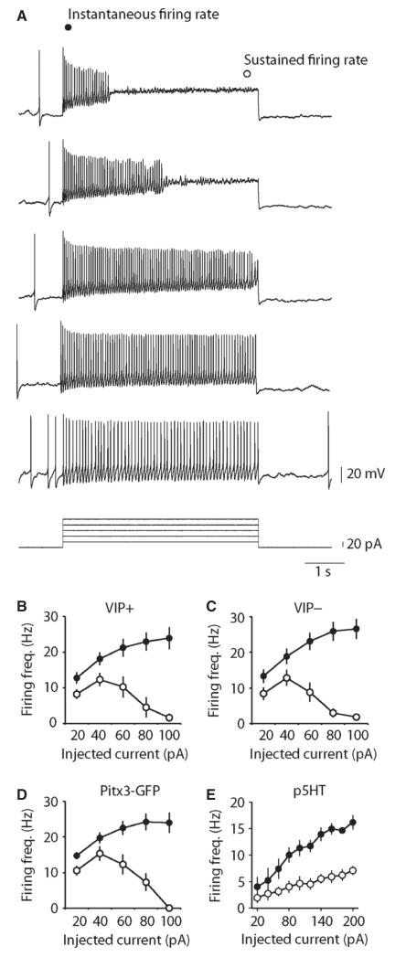
Fig. 3.
DRN/vlPAG dopamine neurons exhibit depolarization-induced spike-frequency adaptation. (A) Example of the effects of depolarizing pulses on AP firing in an identified Pitx3-GFP dopamine neuron. Graphs showing group average responses to depolarizing pulses in TH-GFP/VIP+ (n = 10) (B), TH-GFP/VIP− (n = 13) (C), Pitx3-GFP+ (not labelled with neurobiotin, n = 16) (D) and putative 5HT (GFP−, n = 5) neurons (E). All three groups of GFP+ neurons exhibit clear depolarization-induced spike-frequency adaptation often leading to depolarization block. In contrast, p5HT neurons exhibit strong spike-frequency adaptation with increasing depolarization, but do not enter depolarization block.

