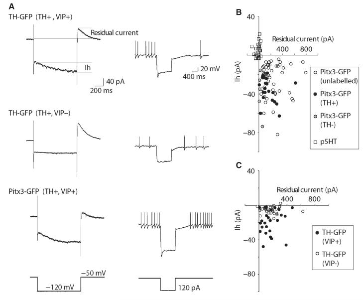
Fig. 4.
DRN/vlPAG dopamine neurons exhibit a hyperpolarization-activated inward current (Ih), which is largest in the VIP+ subgroup. Both groups exhibit an outward residual current following the hyperpolarization that probably contributes to delayed repolarization seen in current clamp. (A) Examples of voltage-clamp (left-hand traces) and current-clamp (right-hand traces) responses to hyperpolarization in different GFP+ neurons. (B) Scatter plot showing Ih and residual current values for Pitx3-GFP+ neurons and p5HT neurons. (C) Scatter plot showing Ih and residual current values for TH-GFP+ neurons neurochemically identified as dopaminergic and either VIP+ or VIP−.

