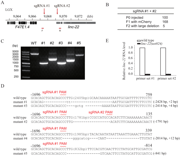
Figure 2. Dual sgRNA-guided deletion of the linc-22 promoter.
(A) Schematic of linc-22 gene. (B) Summary of microinjection experiments. F2 progenies were directly screened by PCR amplification. (C) PCR amplification of the targeted region in the deletion mutants. (D) Sequence alignments of wild-type and mutant animals. Dash indicates deletion. The numbers in parentheses within the sequence represent the number of bases not shown. The number of deleted (−) or inserted (+) bases is indicated on the right of each indel. (E) Quantitative real-time PCR detection of linc-22 expression. Total RNAs were isolated from embryos. eft-3 mRNA was used as an internal control for normalization. N = 3.

