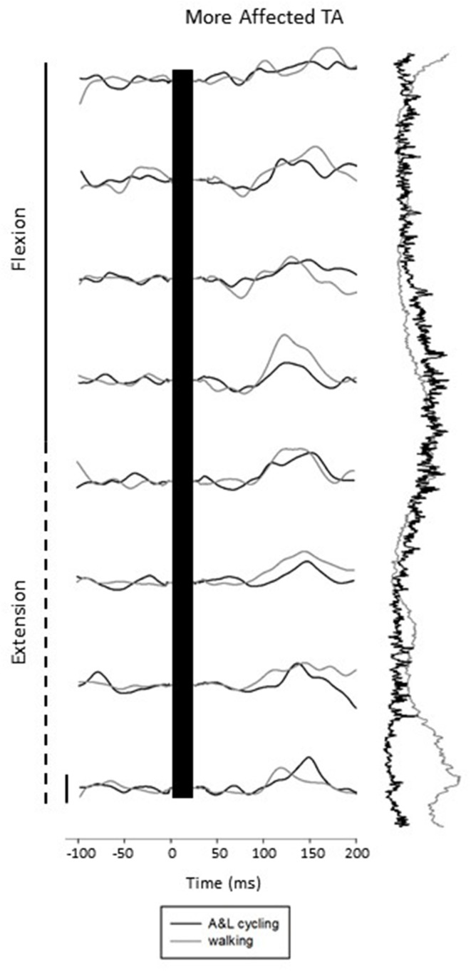
Figure 3.
Subtracted electromyographic (EMG) traces of the more affected tibialis anterior (TA) from a representative participant evoked by superficial radial and superficial peroneal nerve stimulation during A&L cycling and walking. The stimulus artifact has been removed from each trace and replaced by a black bar extending from time 0 out to ~30 ms post stimulus. Background EMG during A&L cycling and walking is shown to the right of the trace plotted vertically. Calibration bar represents 10 μV.

