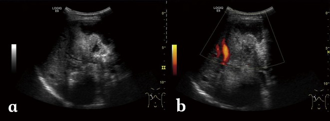
Figure 1.

Alveolar echinococcosis in a 30-year-old woman. (a) Abdominal gray-scale US image shows an irregular type heterogeneous mass lesion with no clear boundary in the right lobe of the liver, containing anechoic pseudo-cystic lesion and hyperechoic foci of calcification. (b) Color Doppler US image shows no obvious blood flow signal while cystic duct is constricted.
