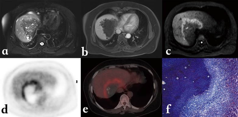
Figure 11.
Alveolar echinococcosis in a 45-year-old patient with hepatic alveolar echinococcosis (HAE). (a) T2-weighted MR images showed a heterogeneous mass: note the multiple vesicles in the lesion, which showed a well-defined border on enhanced T1WI due to the absence of enhancement of the lesion itself (b). (c) Diffusion-weighted MR images revealed a half circular hyper-intensity area at the lesion’s border with the normal liver parenchyma, which was confirmed to be metabolically active by PET (d) and PET/CT (e). (f) HAE peripheral area was composed of severe fibrosis combined with a large number of inflammatory cells on the histopathological sections (Masson staining; ×100).

