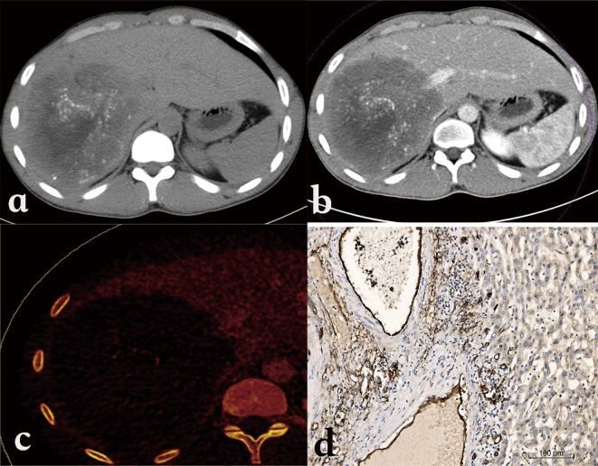
Figure 9.
Alveolar echinococcosis in a 23-year-old woman. (a) Plain CT shows an infiltrative tumor-like mass with irregular margin and heterogeneous density in the right lobe of the liver. (b) Enhanced CT scan shows mild enhancement at the edge of the mass. (c) Iodine map of Spectral CT shows iodine distribution in the liver and lesion. (d) Micro-vessel density on histopathology of the iodine-enhanced area.

