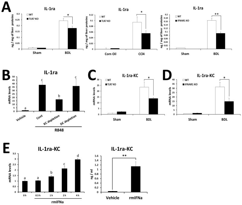
Fig. 5. TLR7- and IFNAR1-deficiency reduce IL-1ra production in KCs.
(A) Hepatic IL-1ra in BDL and CCl4 murine models were quantified by ELISA. The results are expressed as ng of cytokine per mg of liver protein. (B) mRNA of IL-1ra was determined by qRT-PCR in the WT liver at 2 h after injection of TLR7 ligand (R848). Depletion of DCs was achieved by diphtheria toxin injection (4 ng/g) to CD11c-DTR mice for two consecutive days, and depletion of KCs was done by clodronate liposome injection (200μl/mouse) for two consecutive days. (C) mRNA levels of IL-1ra were determined by qRT-PCR in normal KCs, and in vivo activated (21 days post-BDL) KCs from WT and TLR7-deficient mice. (D) mRNA levels of IL-1ra were determined by qRT-PCR in normal KCs, and in vivo activated (21 days post-BDL) KCs from WT and IFNAR1-deficient mice. Values presented are from three independent experiments and each sample was assayed in duplicate. The results are shown as fold change compared with normal cells from WT mice. (E) KCs isolated from WT mice were stimulated with recombinant murine IFNa (500IU/ml), and mRNA and protein levels of IL-1ra were measured with qRT-PCR and ELISA, respectively. Data are presented as means ± SEM per group. Two-tailed Student’s t-test, *P < 0.05, **P < 0.01 (A, C, D). Different letters indicate significant differences (B, p < 0.01; E, p < 0.05) according to ANOVA and DMRT.

