
Figure 7.
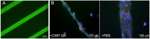
Fluorescence micrographs of PEGDA scaffolds micropatterned with CMP/RGD via surface photopolymerization and further treated with CF-CMP (A), and HUVECs seeded on the micropatterned hydrogels further treated with CMP-QK or blank PBS (B). Width of the line patterns are 200 µm in figure A and 100 μm in B. Cells on CMP-QK modified scaffolds displayed a polarized morphology aligned in the direction of long axis of the micropatterns, while cells on PBS scaffolds exhibited cobblestone morphology. Cells were stained with DAPI (stains nucleus in blue), phalloidin (stains actin in green), and CD31 (stains EC intercellular junctions in red).