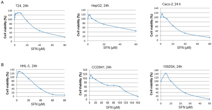
Figure 1. Effects of SFN on the proliferation of normal and tumour cells.
When cells grew to 70–80% confluence, a range of doses of SFN (0–160 µM) were added to the cell culture medium for 24–48 h. The control cells were treated with DMSO (0.1%), and cell viability was determined by the MTT cell proliferation assay (CCD-1092SK cell viability was determined by WST-1 assay according to manufacturer's instructions [88]). Each data point represents the mean ± SD of at least 5 replicates. Statistical significance from the control, *p<0.05, or **p<0.01. A: results from bladder cancer T24, hepatoma HepG2, and colon cancer Caco-2 cells. B: Results from immortalised hepatocyte HHL-5, colon epithelial CCD841, and skin fibroblast CCD-1092SK cell lines.

