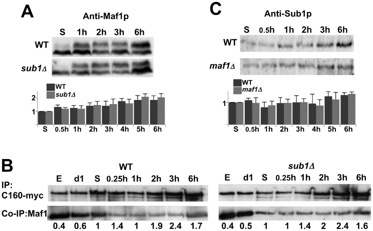
Figure 4. The difference in reactivating Pol III transcription is not due to an excess of Sub1 or Maf1.
The amounts of Maf1 (A) or Sub1 (C) in the indicated strains grown in YPD at day5 or after 1 to 6 h post-inoculation into fresh media were analyzed by SDS-PAGE followed by immunoblotting using polyclonal antibodies as indicated. The signals obtained with anti-Maf1 antibodies correspond to hypo- (fast-migrating) or hyper- (slow migrating) phosphorylated forms of Maf1. The quantifications of Maf1 or Sub1 protein levels were normalized to a control protein band stained by Ponceau Red. Standard deviations were calculated from at least 3 independent experiments. (B) Cells expressing myc-tagged C160, the largest subunit of Pol III, in the presence (WT) or the absence of Sub1 (sub1Δ) were grown in YPD and collected either in exponential phase (E), at day1 (d1) and day5 (S) stationary phase or after 0.25 h to 6 h post-inoculation into fresh medium. Protein extracts were incubated with magnetic beads coated with anti-myc. The bound polypeptides were eluted and analyzed by SDS-PAGE followed by immunoblotting using anti-myc or polyclonal antibodies directed to Maf1. The quantified ratios of co-immunopurified Maf1/immunopurified C160-myc are indicated below each lane.

